This product contains natural rubber latex which may cause allergic reactions.
For health reasons we are unable to accept returns or exchanges of hygiene products.
This item is considered dimensionally oversized parcel or requires truck delivery. Additional shipping charges will be calculated at time of order.
This item is classified as hazardous materials. Can ship ground only - restricted from air. Additional shipping surcharge will be added at time of order.
This product has a Bariatric rating.
If you have any questions please contact customer support at: 03448 730 035 or Email at: customersupport@performancehealth.com
Log in to view when this item will be in stock & ready to ship.
The Estimated ship date is based on shipping address and product quantity.
Estimated Ship Date:
View when this item will be available to ship to your location: Click here
The Muscle Chart Series comprise of a set of 4 unique A1 size charts (33 x 23) that are ideal for Schools, Colleges, Physiotherapy Departments, Hospital Orthopaedic Departments and private Physiotherapy Practices. Two charts feature the major muscles of the front and back of the body. The muscles...
More Info
The Muscle Chart Series comprise of a set of 4 unique A1 size charts (33 x 23) that are ideal for Schools, Colleges, Physiotherapy Departments, Hospital Orthopaedic Departments and private Physiotherapy Practices. Two charts feature the major muscles of the front and back of the body. The muscles are clearly identified and names together with simple explanations defining muscle action and their link to sporting activity as well as exercises for strengthening the muscle. On each of these charts is ADL information (Activities of Daily Living) which helps to better understand muscle function. Superficial muscles are shown in bright red whilst the deep muscles are illustrated in dark red. The two Muscle Attachment charts show and name the Skeletal System as well as where the major muscles are attached on the bone structure. Origins (in red) and Insertions (in blue) are clearly shown and their attachment is described at the side of the chart. This Colour system is used to explain Distal and Proximal Attachments for each muscle.
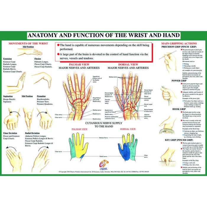
Learning where muscles attach onto the bones help understand how a muscle or group of muscles work to produce body movement. The Joint Anatomy Series comprises 9 x A2 size charts (23 x 16) which are beautifully illustrated in full colour and are encapsulated in plastic film for durability. The following joints: Ankle & Foot, Knee, Hip, Hand & Wrist, Elbow, Shoulder & Spine show the major anatomical structures; including bones, ligaments, tendons and muscles. Each chart provides information about Activities of Daily Living making them ideal for Physiotherapists, Osteopaths, Chiropractors as well as Schools, Colleges and Hospital Orthopaedic Departments. The charts can be purchased individually or as a set of 9 mounted on a calendar for easy hanging on the wall. Chartex Charts are designed with knowledgeable Specialists in the field of medicine (orthopaedics and physiotherapy) in order to set high standards of presentation, helping you to project a professional image.
The Muscle Chart Series comprise of a set of 4 unique A1 size charts (33 x 23) that are ideal for Schools, Colleges, Physiotherapy Departments, Hospital Orthopaedic Departments and private Physiotherapy Practices. Two charts feature the major muscles of the front and back of the body. The muscles are clearly identified and names together with simple explanations defining muscle action and their link to sporting activity as well as exercises for strengthening the muscle. On each of these charts is ADL information (Activities of Daily Living) which helps to better understand muscle function. Superficial muscles are shown in bright red whilst the deep muscles are illustrated in dark red. The two Muscle Attachment charts show and name the Skeletal System as well as where the major muscles are attached on the bone structure. Origins (in red) and Insertions (in blue) are clearly shown and their attachment is described at the side of the chart. This Colour system is used to explain Distal and Proximal Attachments for each muscle.
Learning where muscles attach onto the bones help understand how a muscle or group of muscles work to produce body movement. The Joint Anatomy Series comprises 9 x A2 size charts (23 x 16) which are beautifully illustrated in full colour and are encapsulated in plastic film for durability. The following joints: Ankle & Foot, Knee, Hip, Hand & Wrist, Elbow, Shoulder & Spine show the major anatomical structures; including bones, ligaments, tendons and muscles. Each chart provides information about Activities of Daily Living making them ideal for Physiotherapists, Osteopaths, Chiropractors as well as Schools, Colleges and Hospital Orthopaedic Departments. The charts can be purchased individually or as a set of 9 mounted on a calendar for easy hanging on the wall. Chartex Charts are designed with knowledgeable Specialists in the field of medicine (orthopaedics and physiotherapy) in order to set high standards of presentation, helping you to project a professional image.
More Info
The Muscle Chart Series comprise of a set of 4 unique A1 size charts (33 x 23) that are ideal for Schools, Colleges, Physiotherapy Departments, Hospital Orthopaedic Departments and private Physiotherapy Practices. Two charts feature the major muscles of the front and back of the body. The muscles are clearly identified and names together with simple explanations defining muscle action and their link to sporting activity as well as exercises for strengthening the muscle. On each of these charts is ADL information (Activities of Daily Living) which helps to better understand muscle function. Superficial muscles are shown in bright red whilst the deep muscles are illustrated in dark red. The two Muscle Attachment charts show and name the Skeletal System as well as where the major muscles are attached on the bone structure. Origins (in red) and Insertions (in blue) are clearly shown and their attachment is described at the side of the chart. This Colour system is used to explain Distal and Proximal Attachments for each muscle.
Learning where muscles attach onto the bones help understand how a muscle or group of muscles work to produce body movement. The Joint Anatomy Series comprises 9 x A2 size charts (23 x 16) which are beautifully illustrated in full colour and are encapsulated in plastic film for durability. The following joints: Ankle & Foot, Knee, Hip, Hand & Wrist, Elbow, Shoulder & Spine show the major anatomical structures; including bones, ligaments, tendons and muscles. Each chart provides information about Activities of Daily Living making them ideal for Physiotherapists, Osteopaths, Chiropractors as well as Schools, Colleges and Hospital Orthopaedic Departments. The charts can be purchased individually or as a set of 9 mounted on a calendar for easy hanging on the wall. Chartex Charts are designed with knowledgeable Specialists in the field of medicine (orthopaedics and physiotherapy) in order to set high standards of presentation, helping you to project a professional image.


 US
US France
 Australia